Send me the case, and I send you the patient back.
Referrals are single-tooth cases. Nothing else is examined, nothing else is quoted, and the patient goes back to your chair for everything that follows.
My promise to you“We guarantee the return of the patient after treating that tooth only.”
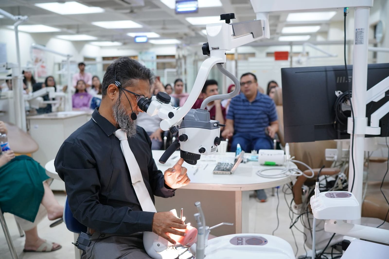
Demonstrating separated-file retrieval at the microscope on a hands-on workshop.
What to refer. Three kinds of case, kept separate.
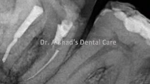
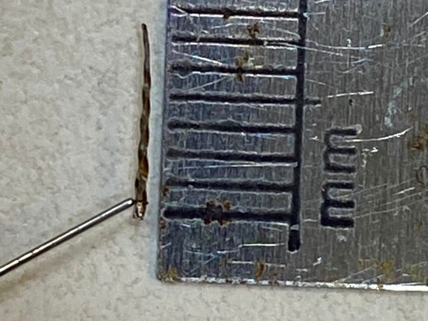
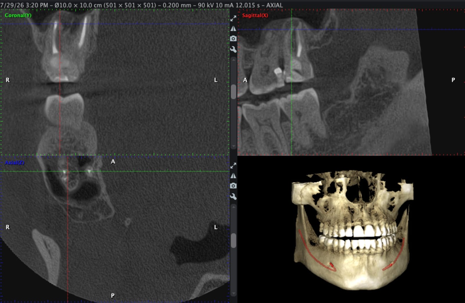
Every film below is shown at or under its true size.
Separated instruments in any canal.
“A small file broken in the upper part of a root canal in a front tooth is fairly simple and straightforward. A difficult case is a long file broken in the mesial root of a second molar, in a patient with limited mouth opening.”
Why these cases come to me.
“The ability to remove a broken or separated instrument comes from three things: training, equipment and experience. I am trained in separated instrument removal and in use of the microscope at the very high magnifications that retrieval requires. Apart from the microscope I have a wide variety of equipment for different challenging cases. I have experience of removing 50+ cases to date.”
“A general dentist does not have the training, the equipment or the experience for handling such cases. I have extensive training and experience over two decades, and almost a decade of using the microscope.”
Cone beam CT is not in the building. When a case needs a 3D scan, I prescribe it to an outside laboratory.
How a referral runs.
- 1
Send it on WhatsApp.
The radiograph and a line about the tooth is enough for me to say whether it is retrievable.
- 2
The patient is seen for that tooth.
Under the microscope, under rubber dam, and nothing outside the referred tooth is treated.
- 3
You get the films and the findings.
What I found, what I did, and what the tooth now needs from you.
- 4
The patient returns to you.
“We guarantee the return of the patient after treating that tooth only.”
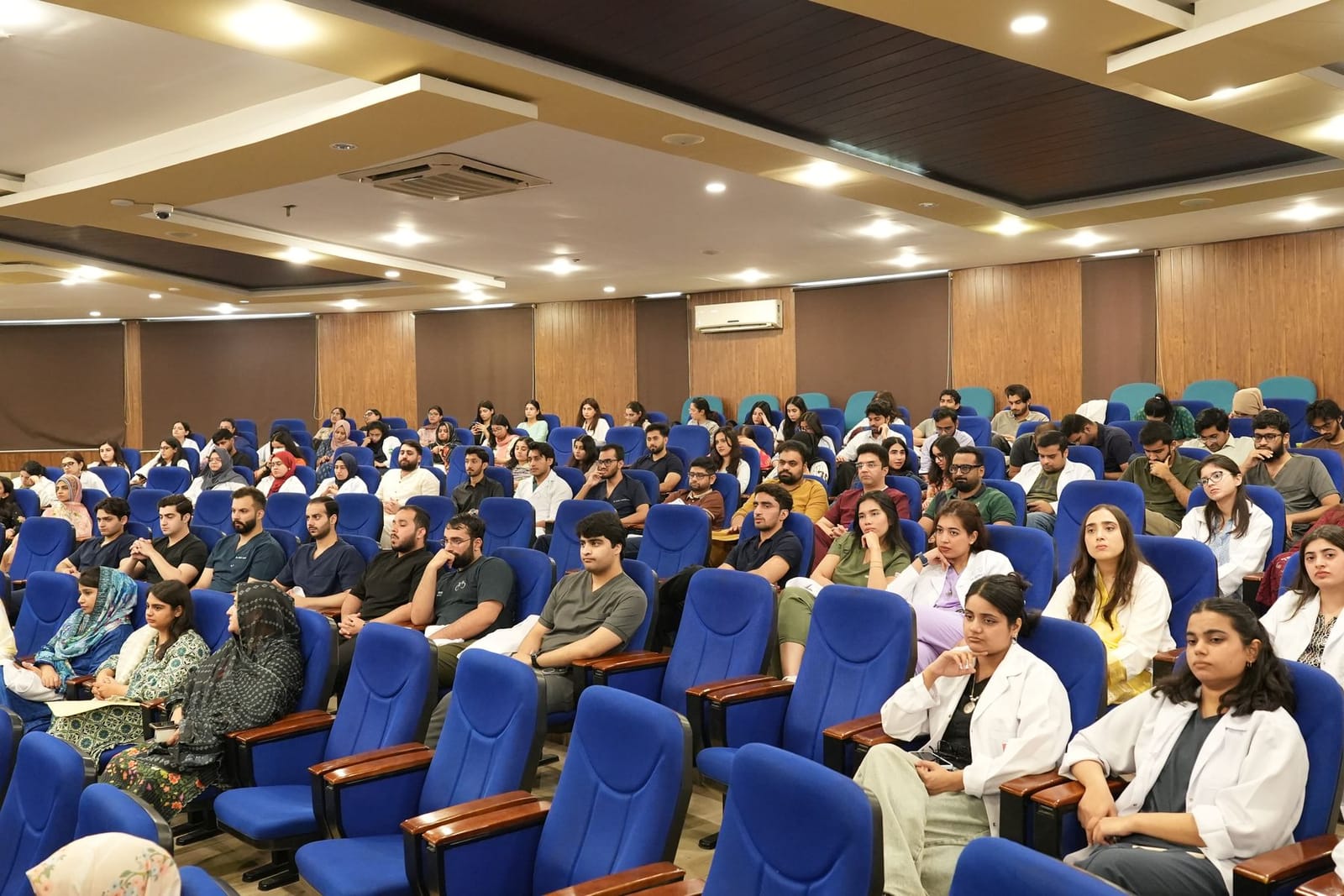
I teach this procedure.
- 300+
- dentists trained in separated-file retrieval
- Nine
- hands-on workshops since 3 May 2024
- 57+
- invited lectures, including “Separated File Removal: Myths & Misconceptions” and “Microscope Enhanced Restorative Dentistry”
Founding Vice President, Pakistan Association of Restorative Dentistry
I review for nine journals, unpaid and by invitation.
- Journal of Endodontics
- Dental Traumatology
- Journal of Visualized Experiments
- BMC Oral Health
- Clinical Oral Investigations
- PLOS One
- Journal of Oral Rehabilitation
- European Journal of Dentistry
- Medicina
Also Honorary Editor, Journal of the Pakistan Dental Association.
16 of my 48 papers are tagged to root canal treatment, re-treatment or separated instruments.
See the full record →Send me the radiograph and a line about the tooth.
Mon–Thu & Sat, 4:00–8:00 PM. Closed Friday & Sunday.